HomeOur laboratoriesImplant Structure Testing Lab

Implant Structure Testing Lab

Microscopic examination:

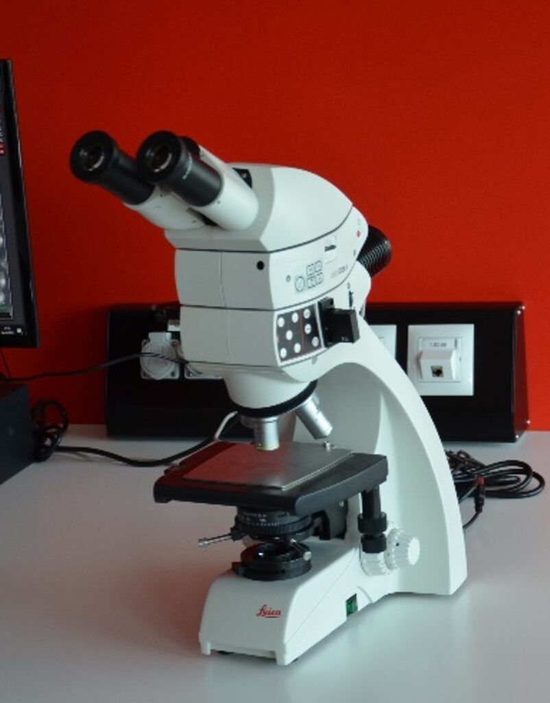
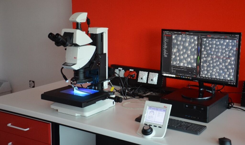
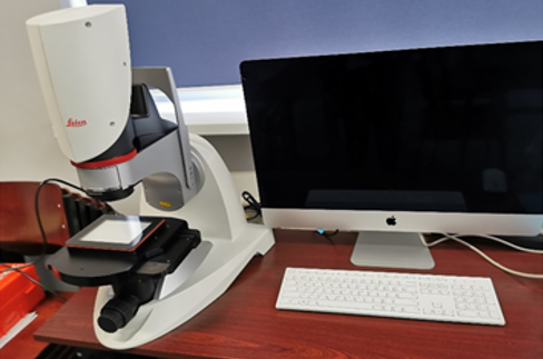
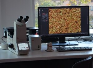
Leica DMi8  microscope
Objectives: 5 x, 10 x, 20 x, 50 x, 100 x
Eyepiece: 10 x

Microscope in an inverted configuration allowing observation of metallographic specimens together with a high resolution digital camera. Fully automated microscope with software and touch panel control and automatic XYZ axis image folding.

Lab supervisor

Anita Kajzer

Anita Kajzer, PhD, Eng.

Anita.Kajzer@polsl.pl
room 110, Zabrze

Leica DM750M microscope
Objectives: 5 x, 10 x, 20 x, 50 x
Eyepiece: 10 x

Metallographic microscope for observing metallographic specimens in conjunction with a high-resolution digital camera.

  • ability to observe specimens in incident and transmitted light for bright field, side illumination and polarisation techniques
  • possibility to record data on SD card
  • ability to control the camera operation and image acquisition  from another workstation via WiFi communication

Modular stereo microscope Leica M205 A
Magnification range: 7.8 x – 160 x (1.0 x objective, 10 x eyepiece)

Parameter:

  • a software module to measure parameters such as area (circle, square, rectangle and any shape), radius, diameter, angle and generate reports of these measurements as an Excel files
  • a software module for generating, visualising and measuring 3D surfaces derived from the scanning an image with extended depth of field in the z-axis. 

Leica S9i stereo microscope
Magnification range: 7.8 x – 160 x (1.0 x objective, 10 x eyepiece)

  • manual Z-axis stand
  • computer and software integration
  • software module for measuring parameters such as area (circle, square, rectangle and any shape), radius, diameter, angle. 

Digital microscope Leica DVM6 A
3 objectives (with magnification range from 12 x to 2350 x)

For real-time inspection, with image capture, video recording and measurement capabilities. Fully automated microscope allowing software control and automatic image assembly in XYZ axes – creating 3D maps of the surface.

The system is equipped with: 

  • tilting head (from -60° to +60°), 
  • field of view width: 43.75 mm, 12.55 mm, 3.6 mm 
  • transmitted and incident light illumination
  • fully motorised stage and all-in-one PC with LAS X software for 2D and 3D analysis
  • motorised axial movement with manual focus.

Surface topography, wetting angle and surface free energy test stations:

Optical tensiometer with Theta 3D topography system

Measurment ranges:

  • wetting angle: 0-180°, accuracy ±0.1°,
  • surface/interphase tension: 0.01 – 1000 mN/m, accuracy ±0.01 mN/m.
  • integrated topograpy module for 2D and 3D surface imaging with the option to correct for surface irregularities in the wetting angle;
  • automated wetting angle testing precisely on the surface with the determined topographic characteristics.

Parameter analysis according to ISO 4287, ISO 4288:

  • r (Wenzel equation)
  • ϴc, wetting angle with roughness correction/Wenzel
  • wetting angle
  • Sdr (%), Sa (um), Sq (um)
  • Horizontal and vertical parameters for any 2D line from the graph Ra, Rq,Rp, Rv, Rz, R10z

Image options:

  • Optical image, 2D and 3D roughness map:
  • Maximum specimen size on stage: unlimited x 180 mm x 22 mm (L x W x H).

Silesian Center for Engineering Support
in Medicine and Sport "Assist Med. Sport Silesia"

Social Media
Contact

European HealthTech Innovation Center

© 2024 EHTIC. All rights reserved